When it comes to keeping your vascular system safe, recognizing the signs of Deep Vein Thrombosis (DVT) early and acting fast can be the difference between normal recovery and life-threatening complications. As a specialist in the field, Dr. Aakash Patel focuses on advanced vascular diagnostics and treatment pathways. In this full guide we will explore what deep vein thrombosis looks like, why timely diagnosis and treatment matter, how advanced imaging such as Venography (also called venogram) supports high-risk cases, and how patients in Ahmedabad and elsewhere can access targeted care. This resource will help you recognize substantial risk indicators, understand how venography functions as a tool for deep thrombosis treatment, and learn why seeking expert care for deep vein thrombosis treatment in Ahmedabad is vital.
Understanding Deep Vein Thrombosis (DVT)
What is DVT?
Deep vein thrombosis refers to the formation of a blood clot in one of the major deep veins of the body — most commonly the legs. This clot can block blood flow, cause swelling and pain, and most critically, it can break off and travel to the lungs, causing a Pulmonary Embolism (PE).
Why it matters
Without appropriate deep vein thrombosis treatment, the risks escalate. Standard therapy prevents clot growth, stops migration of the clot, and reduces chances of recurrence. Delayed care can result in lung complications, long-term venous damage and reduced quality of life.
Who is at risk?
Although anyone can develop DVT, typical risk factors include prolonged immobility (such as long flights or bed rest), recent surgery, trauma, certain cancers, hormone therapy, and inherited clotting disorders. The incidence increases with age and certain anatomical variants.
Recognizing the Signs of DVT
Identifying DVT early — and starting deep thrombosis treatment quickly — improves outcomes and reduces the chance of progression to pulmonary embolism. Some of the major signs include:
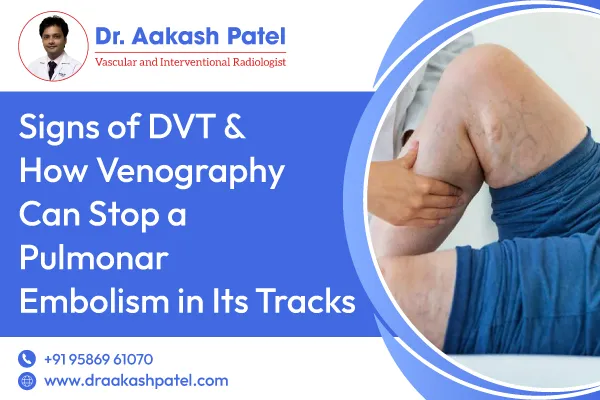
- Swelling in one leg (or arm) is often the calf or thigh.
- Pain or tenderness, particularly when standing or walking.
- Skin discoloration — redness, warmth, or bluish-tinged skin.
- There is a noticeable difference in size between limbs.
- Visible surface veins.
- In more severe cases: shortness of breath, chest pain, rapid heartbeat (if PE occurs).
Because these symptoms can resemble other issues (like muscle injury or varicose veins), medical imaging is often required for definitive diagnosis.
When Standard Tests Fall Short: Why Venography Has a Role
Common testing methods
For most suspected DVT cases, non-invasive tests are used first:
- Blood test (D-dimer) to detect clot-fragment release.
- Duplex ultrasound imaging of leg veins, standard and widely available.
These tools work well in many patients, but they have limitations: ultrasound may not clearly visualize pelvic or deep iliac veins, or the extent of clot burden may be unclear.
What is Venography?
Venography (contrast venogram) is an invasive imaging procedure in which a dye (contrast) is injected into a vein, and X-ray/fluoroscopy captures real-time images of venous flow and blockages. It remains the “gold standard” in certain complex or equivocal cases of DVT.
How Venography helps stop pulmonary embolism
When ultrasound is inconclusive or when a clot is suspected in deep pelvic veins, venography provides precise visualization of the thrombus, its size, location and extension into more proximal veins. That enables targeted intervention — catheter-based clot removal, directed anticoagulation, or filter placement — thereby reducing the risk of the clot migrating and causing pulmonary embolism.
When doctors use it
Venography is often reserved for:
- Patients with suspected DVT but negative or inconclusive ultrasound.
- Clinically high-risk patients (history of PE, severe symptoms) need detailed mapping of clot.
- Pre-procedure planning for thrombectomy or stent placement in cases of complex venous anatomy.
Deep Vein Thrombosis Treatment & Role of Venography
Goals of DVT treatment
For effective deep vein thrombosis treatment, the aims are to:
- Prevent the blood clot from growing.
- Prevent the clot from breaking free and causing pulmonary embolism.
- Reduce the risk of future clot formation.
Standard treatment options
Often the first line of treatment includes anticoagulant medications (“blood thinners”), which help prevent the clot from growing and new clots from forming. For patients with large clots or those at high risk of PE, more aggressive interventions may be used.
When Venography-Guided Treatment Comes In
If venography reveals a significant thrombus burden, especially in pelvic or iliac veins, then treatment may include:
- Catheter-directed thrombolysis (dissolving the clot with medication delivered directly).
- Mechanical thrombectomy (physically removing the clot).
- Placement of an inferior vena cava (IVC) filter in cases when anticoagulation is contraindicated.
By visualizing the clot via venography beforehand, the treatment is more precise, the risk of clot migration reduces, and chances of pulmonary embolism drop significantly.
Deep Vein Thrombosis Treatment in Ahmedabad: Accessing Expert Care
If you or a loved one in Ahmedabad notices persistent leg swelling, tenderness, warmth or skin-changes — this may signal DVT and require prompt assessment. At the clinic of Dr. Aakash Patel in Ahmedabad, you can access diagnostic evaluation including advanced imaging and venography for complex cases. Whether you’re searching for “deep thrombosis treatment near me” or specifically “deep vein thrombosis treatment in Ahmedabad”, it is crucial to partner with a vascular specialist who understands the full spectrum of DVT and PE risk.
By utilizing venography where standard tests fall short, you access advanced diagnostics. That empowers faster, more tailored treatment strategies designed to prevent major complications such as pulmonary embolism. In other words: the combination of early recognition of symptoms, prompt assessment, and advanced imaging gives your care the best possible chance for success.
Recognizing When to Seek Venography Consultation
You should discuss venography-based evaluation with your specialist if:
- You have confirmed or strongly suspected deep vein thrombosis and ultrasound was inconclusive.
- You present symptoms affecting pelvic or iliac veins (deep leg, hip region) where ultrasound may not be sufficient.
- You experienced previous DVT or PE and are at high risk of recurrence.
- You are unable to tolerate anticoagulation and need an alternative evaluation plan.
By bringing these risk factors to your specialist, you can evaluate if venography is appropriate.
Preventing Pulmonary Embolism: The Big Picture
A pulmonary embolism occurs when a blood clot (often from the leg) travels to the lungs, posing a serious threat. Effective deep vein thrombosis treatment prevents clot growth and migration, and applying advanced imaging techniques like venography adds another layer of safety in complex cases.
Lifestyle adjustments also play a key role: keeping active, avoiding prolonged immobility (such as long travel or bed rest), using compression stockings when advised, and following anticoagulant therapy closely. Early detection of deep vein thrombosis (DVT), combined with targeted treatment and advanced imaging, reduces your risk of a pulmonary embolism and supports full recovery.
Why Choose Us
I am Dr. Aakash Patel, specializing in vascular medicine and advanced vein diagnostics. At our clinic in Ahmedabad, we deliver full spectrum care for deep vein thrombosis treatment, including high-level imaging such as venography for complex cases. With our patient-centered approach, advanced technology and personalized plan, we are committed to rapid assessment, precise diagnosis and tailored therapy. Whether you require standard DVT care or advanced venography treatment for DVT, our goal is to help you regain vascular health and avoid serious complications.
Conclusion
When you recognize the signs of deep vein thrombosis, act. Prompt diagnosis and well-chosen deep thrombosis treatment reduce the risk of a clot breaking free and causing pulmonary embolism. While standard tests like ultrasound handle most cases, advanced imaging such as venography treatment for DVT becomes crucial in complex scenarios or when symptoms persist. For those looking for “deep thrombosis treatment near me” or “deep vein thrombosis treatment in Ahmedabad”, partnering with a specialist makes all the difference. Early intervention and precise imaging protect your health, support recovery and safeguard against long-term complications.
For expert evaluation of deep vein thrombosis (DVT) and advanced venography-based treatment, call +91 9586961070 or Book Your Appointment Today for personal consultation.