Table of Contents
- Introduction
Key Takeaways
- Venography provides gold-standard imaging for deep vein thrombosis (DVT), visualizing clots in hard-to-reach deep veins.
- It doubles as a treatment gateway, allowing catheter-based deep thrombosis treatment in one session.
- Common deep vein thrombosis symptoms include leg pain, swelling, warmth, and skin changes demanding urgent evaluation.
- Deep vein thrombosis DVT treatment post-venography focuses on clot resolution and flow restoration via medications or procedures.
- Outpatient venography by Dr. Aakash Patel minimizes recovery time while maximizing diagnostic and therapeutic precision.
Deep vein thrombosis (DVT) strikes silently in many cases, forming blood clots in the deep veins of the legs that can lead to swelling, pain, and life-threatening complications if a clot breaks loose. This procedure empowers precise visualization of these hidden threats, guiding doctors like Dr. Aakash Patel to confirm deep vein thrombosis and initiate targeted deep thrombosis treatment or deep vein thrombosis DVT treatment. For patients, professionals, and families seeking reliable facts on vascular health, this guide breaks down venography's role in modern care.
What Is Deep Vein Thrombosis (DVT)?
Deep vein thrombosis occurs when a blood clot forms in a deep vein, most commonly in the calf or thigh, disrupting normal blood flow back to the heart. These clots develop from factors like prolonged sitting during long flights, recent surgery, immobility from illness, pregnancy, or inherited clotting disorders that make blood more prone to coagulation. Without intervention, the clot can grow, cause tissue damage from poor circulation, or dislodge as a pulmonary embolism, blocking lung arteries and straining breathing or heart function.
The condition often affects one leg, creating an imbalance that patients notice first through localized changes. Medical teams prioritize early detection because timely deep thrombosis treatment can prevent escalation, restore circulation and reduce recurrence risk. Dr. Aakash Patel frequently sees cases where lifestyle demands, such as desk jobs or travel, contribute to this vascular challenge, making awareness critical for at-risk groups.
Common Symptoms of Deep Vein Thrombosis
Patients with deep vein thrombosis (DVT) report distinct signs that signal vascular obstruction. Throbbing pain arises in the calf or thigh, intensifying with walking or standing, as the clot compresses surrounding tissues and nerves. Swelling builds in the affected leg, creating a noticeable size difference that persists despite elevation.
Skin over the area feels warm to the touch, reflecting inflammation from slowed blood flow. Redness or darkened discoloration appears, though it may blend into brown or black skin tones, requiring careful inspection. Veins near the surface become swollen, hard, or tender under pressure, alerting clinicians to deeper issues. These symptoms demand prompt evaluation to differentiate deep vein thrombosis from muscle strains or infections.
The Role of Venography in Diagnosing DVT
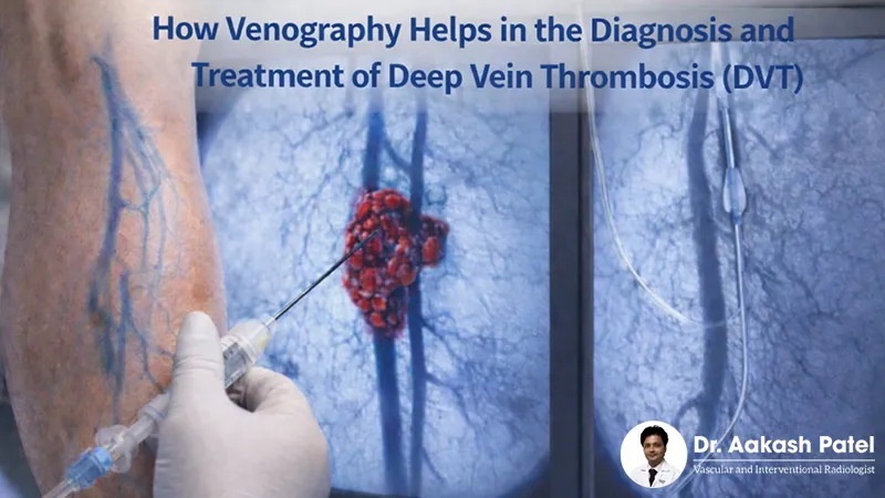
Venography's the go-to test when ultrasounds aren't quite clear enough or when it comes to planning bigger interventions - it's the gold standard for confirming deep vein thrombosis (DVT). This procedure works by injecting a contrast dye into the vein which lets you see in real-time exactly how blood flow is being blocked by clots and where they are in the vein. For Dr. Aakash Patel, venography's the way to map out the full extent of the clot, see if the vein is still open, and identify any blockages that might be influencing the deep vein thrombosis DVT treatment approach.
The dye highlights those areas where the clot is interrupting flow, which is super useful for figuring out whether it's a new clot or an old one. This gives Dr. Patel and his team the precision they need when it comes to deciding between thrombolysis, stenting, or anticoagulation, and they can tailor deep thrombosis treatment to the specific patient's anatomy.
In cases where people have had DVT before, venography is especially helpful - it can reveal underlying vein problems, whether it's compression or scarring, and that information is key to long-term management.
Why Venography Trumps Routine Tests
Of course, ultrasound is the first port of call for deep vein thrombosis, but when it comes to the pelvic or abdominal veins, venography's the way to go. It can spot non-occlusive clots that are only partially blocking flow, which ultrasounds might miss, and it evaluates the function of the valves in the vein - which is super important for preventing post-thrombotic syndrome. For Dr. Patel's patients, this test really bridges the gap when it comes to diagnosis, especially before procedures like venous angioplasty.
How Venography Guides Deep Thrombosis Treatment: The Benefits of Quick & Accurate Diagnosis
So once deep vein thrombosis (DVT) shows up on an image, venography comes in as a bit of a hybrid - it's not just a diagnostic tool, it's also a therapeutic one. The dye injection catheter lets Dr. Patel or his team directly access the clot, which means they can do a mechanical thrombectomy or get medication in to start dissolving the blockage - all in one go. This really cuts down on hospital stays and the potential for complications.
In venous angioplasty situations, venography helps figure out if there's stenosis after the clot's gone, which is where it comes in handy for balloon expansion or stent placement to open up the narrowed segments. And that's all part of an approach that helps restore normal flow dynamics, which reduces the pressure that brings on ongoing symptoms. It all adds up to a level of integrated care that really informs the deep vein thrombosis DVT treatment plan, and sometimes even gets things sorted out on an outpatient basis.
Step-by-Step Venography Procedure
Dr Aakash Patel runs venography as a pretty straightforward outpatient process, usually done under local anaesthetic. Patients turn up, get into a gown, and lie down on an exam table that's all set up with easy access to their legs. They get wrapped up in some protective gear to shield them from any radiation while the team gets everything ready to go.
A local anaesthetic is used to numb the skin - often near where they're putting the catheter in, which is usually either near the foot or the groin. Dr Patel then inserts a catheter under some X-ray guidance, injecting some dye to make the veins stand out & to highlight any clots in the deep vein thrombosis picture. We get real-time images of any blockages & that tells us exactly what to do next, be it aspirating or dilating.
After the procedure, there's a bit of post-care to deal with, lasting a few hours, and the discharge instructions cover things like using compression, moving about a bit and medications. Any soreness at the injection site fades pretty quickly, so most people can get back to light activities pretty soon after having deep thrombosis treatment.
Risks, Recovery & Long-Term Management After Venography
Venography does come with some minor risks like allergic reactions to the dye or temporary irritation to the vein, but Dr Aakash Patel's got measures in place to keep those risks to a minimum - things like pre-medication and getting plenty of fluids in beforehand.
A bit of bruising & slight bleeding at the puncture site is pretty normal and will clear up in a few days. Infection is super rare, but if it does happen the team will jump on it and get you on antibiotics ASAP.
Aftercare is all about taking it easy, drinking plenty of water to flush the dye out & taking some medication to stabilise clots during deep vein thrombosis DVT treatment. Patients are encouraged to take things easy & get moving gently once they're discharged, then gradually get back to normal routines over the next 24-48 hours. Follow-up checks see how symptoms are doing & whether the vein is still clear using duplex scans.
Long-term, it's all about making some lifestyle changes that complement the medical treatment. Compression stockings help keep things flowing & regular activity helps prevent stasis - especially for people who are prone to recurrent deep vein thrombosis. Dr Patel's got all this worked into a plan that's tailored to each individual, with check-ups and the occasional venography if needed to keep things on track.
Why Choose Us
Dr Aakash Patel's practice stands out because it puts patient care at the very heart of everything it does. It's all about getting the imaging right, so we can diagnose deep vein thrombosis accurately and get the deep thrombosis treatment sorted out with minimal fuss. From the moment you first come in to see us, right up to aftercare and follow-up, we're always keeping you in the loop, so you know exactly what's going on with your deep vein thrombosis DVT treatment. And because we use all the latest minimally invasive techniques, you can be back on your feet in no time - and we'll have a treatment plan in place that fits in with your life, whatever that might look like. We're a team of experts working under Dr Patel's guidance, so you can be sure of getting the best possible care.
Conclusion
Venography really is a gamechanger for deep vein thrombosis (DVT) treatment - it gives us super-clear pictures of exactly what's going on with the clot and the vein, which lets us get the right care in place right away. It's not just a diagnostic tool, but a treatment in its own right too - we can use it to spot problems that might otherwise have gone undetected, and get immediate treatment started, all in the comfort of a day-care setting with a specialist like Dr Aakash Patel at the helm. Patients can get moving faster, without the risks that come with being stuck in one place for too long, and they get the tools they need to prevent further problems in the long run.
Ready for expert venography and deep vein thrombosis solutions? Call Dr. Aakash Patel at +91 95869 61070 or book your appointment to diagnose and treat DVT effectively.
Frequently Asked Questions
venography deep vein thrombosis dvt diagnosis deep thrombosis treatment venous imaging procedure blood clot diagnosis catheter based treatment vascular imaging minimally invasive vascular procedure dvt management